
The Role of Radiology in Medicine: What You Need to Know

Radiology is the branch of medicine that uses imaging technology to diagnose disease, guide treatment, monitor therapy response, and support population-wide screening programs. It sits at the center of nearly every clinical pathway, from a routine chest X-ray to a complex cancer staging workup. The American College of Radiology (ACR), the American Board of Radiology (ABR), and the Radiological Society of North America (RSNA) collectively set the standards that govern how imaging is ordered, performed, interpreted, and communicated across U.S. healthcare. Research published through NIH/PubMed Central confirms that radiology’s contribution extends well beyond image reading: radiologists now participate directly in multidisciplinary care teams, shaping clinical decisions alongside surgeons, oncologists, and primary care physicians.
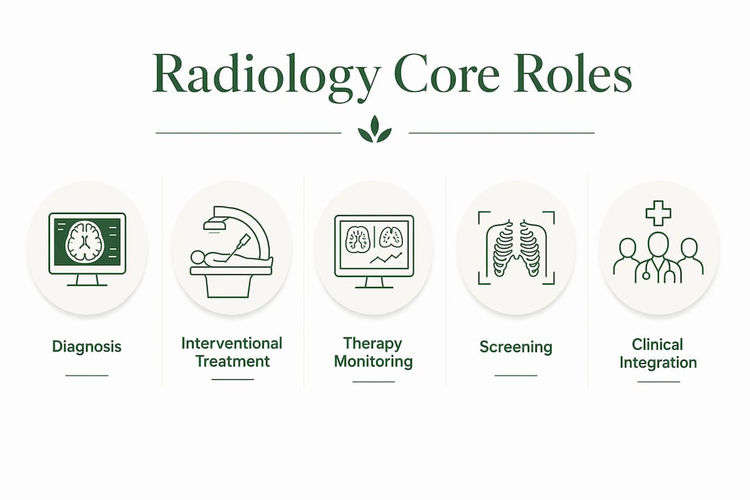
The four core roles imaging plays in modern medicine are:
- Diagnosis: Identifying disease, injury, or abnormality through non-invasive or minimally invasive imaging
- Intervention: Delivering image-guided treatment that often replaces open surgery
- Monitoring: Tracking therapy response and disease progression over time
- Screening: Detecting preclinical disease in defined populations before symptoms appear
Table of Contents
- What does radiology actually cover?
- How radiology shapes diagnosis and clinical decisions
- What interventional radiology can do instead of surgery
- How screening programs use imaging to catch disease early
- How are radiologists trained and certified in the United States?
- How is technology changing what radiologists can do?
- What patients should know about radiation safety and imaging risks
- What peer-reviewed evidence says about radiology’s impact
- How to prepare for imaging and get the most from your results
- Key Takeaways
- Why radiology’s integration into care teams changes everything
- FAQ
What does radiology actually cover?
Radiology divides into two broad areas: diagnostic imaging and interventional radiology. Diagnostic imaging is the information-gathering side, where radiologists interpret images to answer clinical questions. Interventional radiology (IR) is the treatment side, where physicians use real-time imaging to guide instruments through the body and perform procedures with minimal incisions.
The major imaging modalities and their primary uses are:
- X-ray: First-line evaluation of bone fractures, chest infections, and foreign bodies
- Computed Tomography (CT): Rapid cross-sectional imaging for trauma, pulmonary embolism, abdominal emergencies, and cancer staging
- Magnetic Resonance Imaging (MRI): Soft-tissue detail for brain, spine, joints, and pelvic organs, without ionizing radiation
- Ultrasound: Real-time imaging for abdominal organs, obstetric evaluation, vascular flow, and procedure guidance
- Positron Emission Tomography (PET): Metabolic imaging that maps cellular activity, most commonly used in oncology staging and treatment response
- Nuclear medicine: Functional studies such as bone scans, thyroid imaging, and cardiac perfusion tests
- Fluoroscopy: Continuous X-ray used for swallowing studies, joint injections, and catheter placement
A straightforward diagnostic example: a patient with sudden chest pain gets a CT pulmonary angiogram, and the radiologist identifies a bilateral pulmonary embolism within minutes, allowing the emergency team to start anticoagulation immediately. On the interventional side, that same patient’s clot burden might later be managed with catheter-directed thrombolysis, where an IR physician threads a catheter directly into the pulmonary artery under fluoroscopic guidance. Both scenarios rely on the same imaging infrastructure, but the clinical role is entirely different.
How radiology shapes diagnosis and clinical decisions
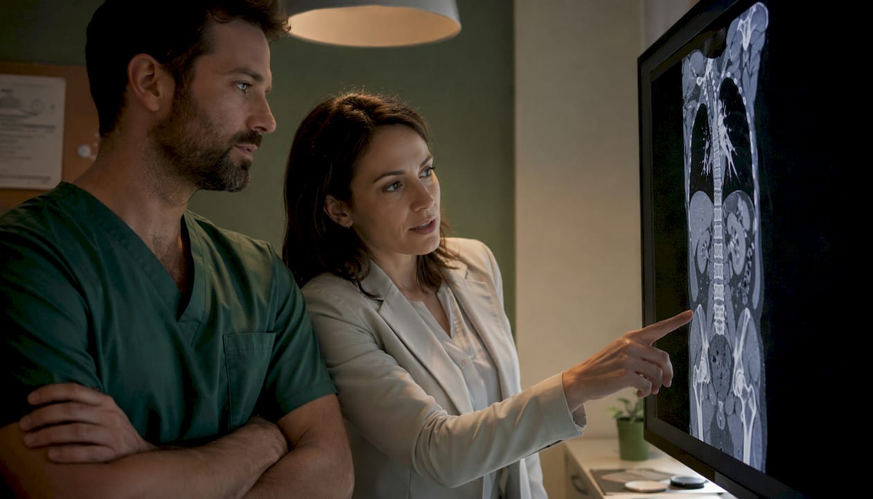
Imaging reduces diagnostic uncertainty in ways that directly change what clinicians do next. A differential diagnosis that might include five or six possibilities often narrows to one or two after a well-chosen scan. That shift in probability is not just intellectually satisfying; it determines whether a patient goes to surgery, gets a biopsy, starts chemotherapy, or goes home with reassurance.
The diagnostic radiology workflow functions as an information-gathering and interpretation activity embedded in the broader diagnostic process, and effective communication between clinical and radiology teams is what makes it work. A radiologist who understands the clinical question can tailor the report to answer it precisely, rather than listing every incidental finding without context.
Imaging answers clinical questions in several concrete ways:
- Rule in: Confirming a suspected diagnosis (e.g., appendicitis on CT)
- Rule out: Excluding a dangerous condition (e.g., no intracranial hemorrhage on CT head)
- Characterize extent: Staging cancer, mapping tumor margins before surgery
- Guide biopsy: Ultrasound or CT-guided needle placement for tissue sampling
- Monitor response: Comparing tumor size before and after chemotherapy cycles
- Direct intervention: Identifying the exact vessel or lesion an IR physician needs to target
In trauma care, CT has become the standard for rapid whole-body assessment. A trauma CT can identify solid organ lacerations, pneumothorax, pelvic fractures, and vascular injuries in a single pass, giving surgeons the information they need before a patient reaches the operating room. In stroke care, CT angiography and perfusion imaging identify salvageable brain tissue and guide the decision to perform mechanical thrombectomy, a time-critical intervention where minutes genuinely matter.
Pro Tip: When your clinician recommends imaging, ask three questions: “What specific finding are you looking for?” “How will the result change my treatment plan?” and “Is there a lower-radiation or non-radiation alternative that answers the same question?” These questions help you understand the purpose of the test and confirm it is the right one for your situation.
What interventional radiology can do instead of surgery
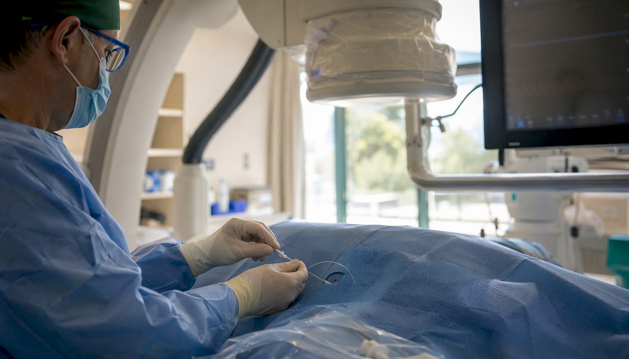
Interventional radiology has grown into a full treatment specialty. IR physicians use real-time imaging, primarily fluoroscopy, ultrasound, and CT, to guide catheters, needles, and other instruments through blood vessels or tissue to treat conditions that once required open surgery. The result is often a shorter hospital stay, less blood loss, faster recovery, and lower complication rates.
| Procedure | Typical Indication | Setting |
|---|---|---|
| Uterine fibroid embolization (UFE) | Symptomatic uterine fibroids | Outpatient |
| Angioplasty and stent placement | Peripheral arterial disease, renal artery stenosis | Inpatient or outpatient |
| CT- or ultrasound-guided biopsy | Tissue sampling for cancer diagnosis | Outpatient |
| Percutaneous drainage | Abscess, pleural effusion, biliary obstruction | Inpatient or outpatient |
| Transarterial chemoembolization (TACE) | Hepatocellular carcinoma | Inpatient |
| Inferior vena cava (IVC) filter placement | High-risk pulmonary embolism prevention | Inpatient |
Consider a patient with a large liver abscess who is too medically fragile for general anesthesia. An IR physician places a drainage catheter under CT guidance in under an hour, the abscess resolves over days with antibiotics, and the patient avoids a laparotomy entirely. That kind of outcome, avoiding major surgery in a high-risk patient, is what makes interventional radiology one of the most consequential specialties in modern hospital care. For a deeper look at how imaging guides treatment beyond diagnosis, the Radiology 101 overview covers common IR procedures and their clinical rationale.
Common benefits of IR over open surgery include:
- Smaller incisions or no incision at all
- Local or moderate sedation instead of general anesthesia
- Same-day or next-day discharge in many cases
- Preserved organ function (e.g., UFE preserves the uterus)
How screening programs use imaging to catch disease early
Early detection through imaging-based screening can improve outcomes by identifying disease before symptoms appear, when treatment is typically less aggressive and more effective. The ACR and other professional bodies publish evidence-based guidelines that define which populations benefit from specific screening programs, and following those guidelines is what separates high-value screening from indiscriminate testing.
Key imaging-based screening programs and their target populations include:
- Mammography: Annual screening recommended for women at average risk starting at age 40 (ACR guideline); detects breast cancer at earlier, more treatable stages
- Low-dose CT (LDCT) for lung cancer: Recommended annually for adults aged 50–80 with a significant smoking history; the U.S. Preventive Services Task Force supports this for high-risk individuals
- Colonoscopy and CT colonography: Colorectal cancer screening options for average-risk adults starting at age 45
- Bone density scan (DEXA): Osteoporosis screening for women 65 and older and younger postmenopausal women with risk factors
- Abdominal aortic aneurysm (AAA) ultrasound: One-time screening for men aged 65–75 who have ever smoked
Imaging-based screening detects preclinical disease and can make treatment less invasive and more effective when targeted to appropriate populations. That qualifier matters. Screening works when it is applied to the right group at the right interval. Applied too broadly, it generates false positives, unnecessary biopsies, and patient anxiety without a net health benefit. The ACR Appropriateness Criteria provide clinicians with evidence-based guidance on which imaging test fits which clinical scenario, reducing the risk of both overuse and underuse.
Screening impact: Low-dose CT screening for lung cancer in high-risk adults has been shown to reduce lung cancer mortality in eligible populations, according to clinical trial data supporting current USPSTF recommendations.
For a practical breakdown of which screenings apply at different ages, the adult screening guide from Gardenstatemedicalgroup is a useful starting point.
How are radiologists trained and certified in the United States?
Radiologists are fully licensed medical doctors. The path to independent practice takes at least 13 years after high school and involves multiple credentialing milestones.
The training timeline runs as follows. Four years of undergraduate education lead to four years of medical school, where students earn an MD or DO degree. After medical school, physicians complete a one-year internship (transitional or preliminary year) that provides broad clinical exposure. They then enter a four-year diagnostic radiology residency, during which they rotate through all major imaging modalities and subspecialties under supervision. Most radiologists complete an additional one- to two-year fellowship in a subspecialty such as neuroradiology, musculoskeletal radiology, interventional radiology, breast imaging, or pediatric radiology.
Board certification through the ABR is the primary credential patients and hospitals look for. The ABR administers a two-part examination process: the Core Exam (taken after 36 months of residency) and the Certifying Exam (taken after residency completion). Certified radiologists must also meet Maintenance of Certification (MOC) requirements to keep their credentials current.
Credentials and affiliations worth knowing:
- ABR certification: Confirms the radiologist passed rigorous written and oral examinations in diagnostic or interventional radiology
- ACR membership or fellowship (FACR): Signals engagement with national standards and continuing education
- Subspecialty fellowship training: Indicates focused expertise in a specific area (e.g., neuroradiology, IR, breast imaging)
- State medical license: Required for independent practice in any U.S. state
- Hospital privileges: Granted by individual institutions after credentialing review
How is technology changing what radiologists can do?
Technology is expanding radiology’s reach while also creating new workflow and quality challenges that the specialty is still working through. The shift is not just about faster machines; it is about who has access to imaging expertise and how that expertise gets applied.
Key technologies and their practical effects:
- Picture Archiving and Communication Systems (PACS): Digital image storage and retrieval that replaced film, allowing instant access to prior studies for comparison and enabling remote reading
- Teleradiology: Remote image interpretation that extends subspecialty expertise to hospitals and clinics that cannot staff a full radiology department around the clock; particularly valuable for overnight and rural coverage
- Artificial intelligence (AI) and decision support: AI tools are now FDA-cleared for tasks including detection of intracranial hemorrhage, pulmonary nodule measurement, and fracture identification; they function as a second reader or triage flag, not a replacement for the radiologist
- Fusion imaging (PET-CT, PET-MRI): Combines metabolic and anatomical data in a single study, improving staging accuracy in oncology and reducing the need for separate scans
- ACR Appropriateness Criteria and clinical decision support (CDS): Electronic tools embedded in ordering systems that check whether a requested study matches evidence-based guidelines before the order is placed
Three near-term impacts worth watching: First, AI triage tools will continue to shorten turnaround times for urgent studies like stroke and trauma CT, getting critical results to clinicians faster. Second, clinical decision support at the point of ordering will reduce low-value imaging by flagging studies unlikely to change management. Third, teleradiology growth, while improving access, risks reducing the face-to-face radiologist-clinician interaction that multidisciplinary care depends on. The specialty’s challenge is preserving that clinical presence even as reading becomes more distributed. For a closer look at where diagnostic imaging is headed, the radiology trends overview covers AI, PACS, and teleradiology in more detail.
What patients should know about radiation safety and imaging risks
Imaging is safe when used appropriately, and the radiology profession has built a substantial infrastructure of protocols and oversight to keep it that way. The central principle is ALARA: As Low As Reasonably Achievable. Every imaging protocol is designed to use the minimum radiation dose that still produces a diagnostic-quality image.
Patient-facing safety measures you should expect:
- ALARA dose optimization: Technologists adjust CT parameters based on patient size and the clinical question, avoiding one-size-fits-all protocols
- Dose reference levels: National benchmarks that facilities compare against to identify when their protocols are delivering higher-than-expected doses
- Contrast screening: Before administering iodinated or gadolinium-based contrast agents, staff screen for kidney function, allergies, and medication interactions (notably metformin with iodinated contrast)
- Informed consent for interventional procedures: IR procedures carry procedural risks beyond radiation; patients receive a formal consent discussion covering benefits, alternatives, and potential complications
- Pregnancy screening: Technologists ask about pregnancy before any study involving ionizing radiation, and protocols exist to minimize fetal exposure when imaging is clinically necessary
- Incidental finding management: Radiologists flag unexpected findings and recommend follow-up imaging or referral, with structured reporting systems (such as ACR’s Lung-RADS and BI-RADS) guiding next steps
Radiologists play a key role in protecting patients from inappropriate radiation exposure by applying dose-optimization strategies and decision-support tools. That responsibility extends to declining or redirecting orders for studies that are unlikely to change management.
Pro Tip: Before any imaging study, ask the technologist: “What is the approximate radiation dose for this exam, and how does it compare to background radiation?” For contrast studies, ask: “What contrast agent will be used, and what should I watch for afterward?” These are standard questions that any imaging team should answer clearly and without hesitation.
What peer-reviewed evidence says about radiology’s impact
Peer-reviewed studies confirm radiology’s central role in healthcare delivery, but they also identify overuse as a source of real harm and unnecessary cost. Both sides of that finding matter.
“High and growing volumes of potentially low-value imaging are a documented concern; imaging that does not answer a specific clinical question can lead to overdiagnosis and patient harm.”
Source: Diagnostic excellence, imaging stewardship, and re-establishing the value proposition in radiology (PMC)
The distinction the evidence draws is precise: high-quality imaging that answers a specific clinical question is high-value care. Indiscriminate imaging with low pre-test probability often causes harm through incidental findings that trigger unnecessary downstream procedures, patient anxiety, and added cost. That is not a reason to avoid imaging; it is a reason to order it thoughtfully.
On the value side, a multi-society expert statement on radiology in value-based healthcare argues that radiology can produce long-term health gains even when short-term imaging costs appear high, and that radiology must be included in value calculations rather than treated as a pure cost center. The same analysis notes that when imaging is ordered during an emergency department visit, average length of stay increases with imaging performed during an emergency department visit, reflecting the time required to perform and interpret studies. That operational reality is a planning consideration, not an argument against imaging; the clinical benefit of an accurate diagnosis in the ED far outweighs the added time in most cases.
The ESR white paper on radiology’s evolving role documents the shift from remote reporting toward central participation in multidisciplinary teams, where radiologists contribute clinical management knowledge alongside image interpretation. That shift has measurable implications for care quality: radiologists who attend tumor boards and trauma conferences influence treatment decisions in ways that a written report alone cannot.
How to prepare for imaging and get the most from your results
Getting the most from a radiology study starts before you arrive at the imaging center. Preparation affects image quality, safety, and how useful the results are to your care team.
Preparation checklist:
- Bring prior imaging studies on CD or confirm they are accessible in a shared electronic system
- Follow fasting or hydration instructions specific to your exam (CT with contrast often requires fasting; MRI of the abdomen may require nothing by mouth for several hours)
- List all medications, especially metformin, blood thinners, and any known contrast allergies
- Arrive with your insurance information and any required prior authorization documentation
- For interventional procedures, arrange a driver; sedation or analgesia is common
Questions that help confirm your imaging will actually influence your care:
- “Is this test likely to change my treatment plan, or is it being ordered to confirm something you already know?”
- “What are the alternatives, including watchful waiting or a different imaging modality?”
- “Will a radiologist’s report come back to you directly, or do I need to follow up to get results?”
- “Does my insurance require prior authorization, and has that been submitted?”
Cost and insurance are practical realities. Many insurers require prior authorization for advanced imaging like MRI and PET. If your clinician uses ACR Appropriateness Criteria or a clinical decision support tool to justify the order, that documentation often speeds the authorization process. When cost is a concern, ask whether a lower-cost modality (ultrasound instead of MRI, for example) answers the same clinical question. For guidance on when a direct radiology consultation makes sense, the when to see a radiologist guide from Gardenstatemedicalgroup walks through the referral pathways clearly.
Key Takeaways
Radiology’s role in medicine spans diagnosis, image-guided treatment, disease monitoring, and population screening, and its value depends entirely on using the right test for the right patient at the right time.
| Point | Details |
|---|---|
| Four core roles | Radiology covers diagnosis, interventional treatment, therapy monitoring, and population screening. |
| Training is extensive | U.S. radiologists complete extensive training, including medical school, internship, residency, and often a fellowship, before ABR certification. |
| Safety is structured | ALARA, dose reference levels, contrast screening, and informed consent protocols protect patients in every imaging encounter. |
| Overuse is a real risk | Imaging that does not answer a specific clinical question can cause harm through incidental findings and unnecessary downstream procedures. |
| Gardenstatemedicalgroup | Offers integrated radiology and diagnostic testing alongside primary care and specialty programs for coordinated, multidisciplinary care. |
Why radiology’s integration into care teams changes everything
There is a version of radiology that most people picture: a physician in a dark room reading films alone, sending a report that arrives hours later. That model still exists in some settings, but it is increasingly the exception rather than the rule, and the difference in patient outcomes between that model and a fully integrated one is significant.
When a radiologist sits at a tumor board alongside the oncologist, surgeon, and pathologist, the imaging interpretation becomes a live conversation. The radiologist can clarify what a finding means in the context of the patient’s history, flag a measurement that changes staging, or recommend a follow-up study that the written report alone might not have prompted. That kind of real-time input is what the medical imaging role in diagnosis and care literature consistently identifies as the highest-value use of radiology expertise.
What I find underappreciated is how much the quality of the clinical question determines the quality of the imaging result. A vague order (“abdominal pain, rule out everything”) produces a vague report. A precise order (“right lower quadrant pain, rule out appendicitis, patient is 28 weeks pregnant”) produces a targeted, actionable answer. Radiologists can only work with the information they are given, which means the clinician-radiologist relationship is genuinely bidirectional. Practices that invest in that relationship, through shared systems, regular communication, and multidisciplinary meetings, get measurably better diagnostic performance than those that treat radiology as a transactional service.
The other thing worth saying plainly: the push toward AI and teleradiology is real and largely positive, but it carries a risk of commoditizing the specialty. Speed and access matter. So does the radiologist who knows your patient’s history, notices that the new finding looks different from last year’s scan, and picks up the phone. Technology should support that relationship, not replace it.
Radiology and diagnostic testing at Gardenstatemedicalgroup
Gardenstatemedicalgroup offers radiology and diagnostic testing as part of an integrated, multidisciplinary practice serving North Bergen and Secaucus, New Jersey. Rather than sending you to a separate imaging center and waiting for results to filter back through disconnected systems, the practice coordinates imaging directly with primary care and specialty services under one roof. That means your clinician has access to your imaging history, your radiologist understands your care context, and follow-up happens without the gaps that come from fragmented care.
If you have been referred for imaging or want to understand which diagnostic tests apply to your situation, contact Gardenstatemedicalgroup to schedule a consultation. This content is for general informational purposes and is not a substitute for professional medical advice; always confirm recommendations with your own clinician.
Authoritative sources and further reading
The sources below are the primary references for the clinical claims in this article. Each serves a distinct purpose.
- American College of Radiology (ACR): The ACR sets clinical practice guidelines, Appropriateness Criteria, and professional standards for U.S. radiologists. Start here for evidence-based imaging recommendations.
- Radiological Society of North America (RSNA): The RSNA publishes peer-reviewed radiology journals and hosts the world’s largest radiology conference; its patient education resources are clear and reliable.
- American Board of Radiology (ABR): The ABR administers board certification examinations and Maintenance of Certification; use this site to verify a radiologist’s credentials.
- NIH/PubMed Central (PMC): Open-access peer-reviewed literature, including the ESR white paper on radiology’s evolving clinical role and the PMC review on imaging stewardship.
- Cleveland Clinic radiology overview: A patient-facing overview of imaging modalities and what to expect; useful for readers preparing for a first scan.
- MedlinePlus imaging and radiology encyclopedia: A plain-language, patient-oriented definition of radiology and its major modalities, maintained by the National Library of Medicine.
- AJR: The role of diagnostic radiology in the diagnostic process: A peer-reviewed article outlining 10 principles for effective information flow between clinical and radiology teams; relevant for clinicians and medical students.
- Multi-society expert statement on radiology in value-based healthcare: A policy-level document from multiple radiology societies arguing for radiology’s inclusion in value-based care calculations; relevant for health systems and policymakers.
FAQ
What is the role of radiology in modern medicine?
Radiology provides the imaging infrastructure for diagnosis, image-guided treatment, therapy monitoring, and population screening across virtually every medical specialty. Radiologists interpret studies, guide interventional procedures, and contribute to multidisciplinary care teams that shape treatment decisions.
How is radiology used in the diagnosis of disease?
Imaging narrows differential diagnoses, confirms or excludes specific conditions, stages disease extent, and guides tissue sampling through CT- or ultrasound-directed biopsy. The diagnostic radiology workflow functions as an information-gathering activity embedded in the broader clinical process, with the radiologist’s report serving as a structured answer to the clinician’s question.
Are radiologists medical doctors?
Yes. Radiologists are fully licensed physicians who complete at least 13 years of training, including undergraduate education, medical school, a one-year internship, a four-year residency, and often a fellowship before earning ABR board certification.
Why does radiology matter for patient safety?
Radiologists apply ALARA dose-optimization principles, screen patients for contrast contraindications, and use clinical decision support tools to prevent inappropriate radiation exposure. They also flag incidental findings and recommend structured follow-up, reducing the risk that an unexpected abnormality goes unaddressed.
When should you ask about imaging appropriateness?
Ask your clinician whether the recommended study is likely to change your treatment plan and whether a lower-radiation or non-radiation alternative answers the same question. ACR Appropriateness Criteria, available to clinicians through electronic decision-support tools, provide evidence-based guidance on which imaging test fits which clinical scenario.
Recommended
- Radiology 101: Beyond the Diagnosis and How It Treats Conditions | Garden State Medical Group | Garden State Medical Group
- The Role of Medical Imaging in Diagnosis and Care
- Radiology Trends: What’s In Store For Diagnostic Imaging? | Garden State Medical Group | Garden State Medical Group
- Our Guide to When You Should See a Radiologist | Garden State Medical Group | Garden State Medical Group